
Linear, curvilinear or phased array?
Choosing the right probe is half of a good scan. The choice is a trade between detail and depth.
Live simulator — try itFull interactive lesson →
The three probes
- Linear (≈5–15 MHz): flat face, rectangular image, fine detail but shallow — vessels, nerves, pleura, needles.
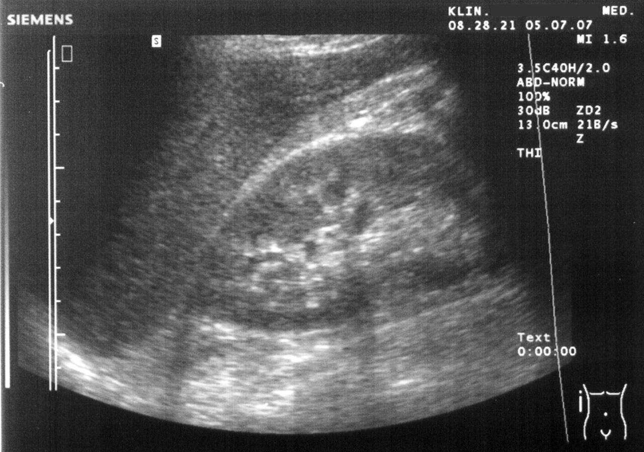
- Curvilinear (≈2–5 MHz): curved face, wide fan, sees deep — abdomen, pelvis, FAST, lung bases.
- Phased array (≈1–5 MHz): tiny footprint that steers its beam between ribs — the heart.
The probe marker
Every probe has a marker on one side, matching a dot on the screen. In emergency and radiology convention the screen dot is on the left: marker to the patient’s head in long axis, to the patient’s right in transverse. Cardiology traditionally flips this.
Real scans
Learn this with your hands. Sonoduck gives you a 3D patient, a probe and a coach that tells you what to do. Free to start, no signup.
Scan your first patientFAQ
Why does high frequency give better detail?
Shorter wavelength and pulse length improve resolution, but higher frequencies attenuate faster, so they don’t reach as deep.
Related guides
- Knobology: the four knobs that matterHow to set ultrasound gain, depth, focus and frequency — with a live simulator so you can turn the knobs and see what happens.
- Ultrasound artifacts, explainedAcoustic shadowing, posterior enhancement, reverberation (A-lines), B-lines, mirror image and edge artifacts — why they happen and how they help diagnosis.
- The FAST exam, explained by actually doing itHow to perform a FAST exam: RUQ, LUQ, pelvic and subxiphoid views, what a positive scan looks like, common pitfalls — with a live ultrasound simulator you can try.
Educational content for learners; follow local protocols and supervision. Simulated images are generated by Sonoduck’s physics model; real images are credited individually.