
The FAST exam, explained by actually doing it
The FAST exam (Focused Assessment with Sonography in Trauma) is a two-minute bedside ultrasound that asks one question: is there free fluid where there shouldn’t be? In an injured, hypotensive patient, free fluid is presumed to be blood.
Live simulator — try itFull interactive lesson →
The four windows
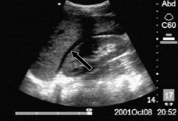
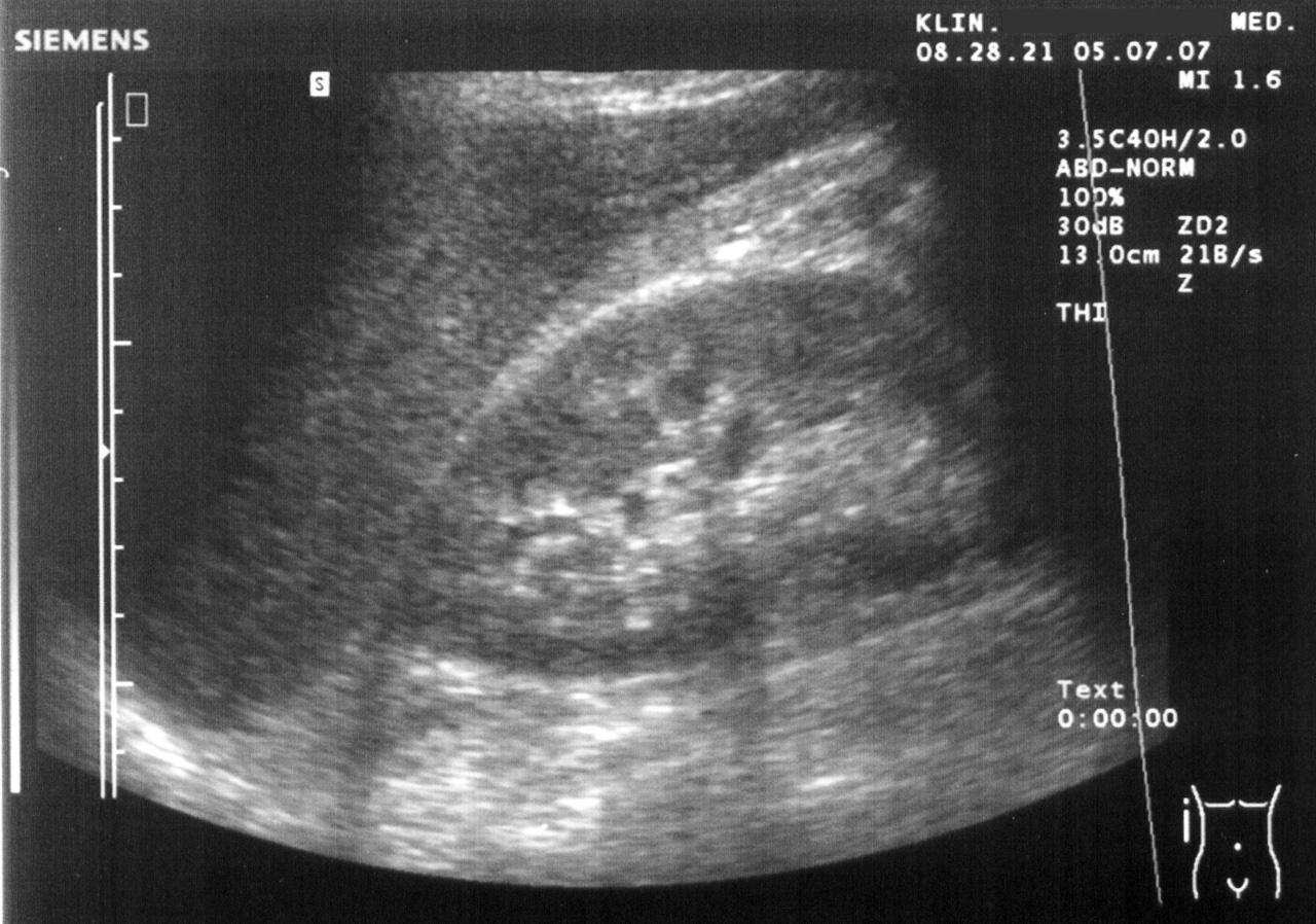
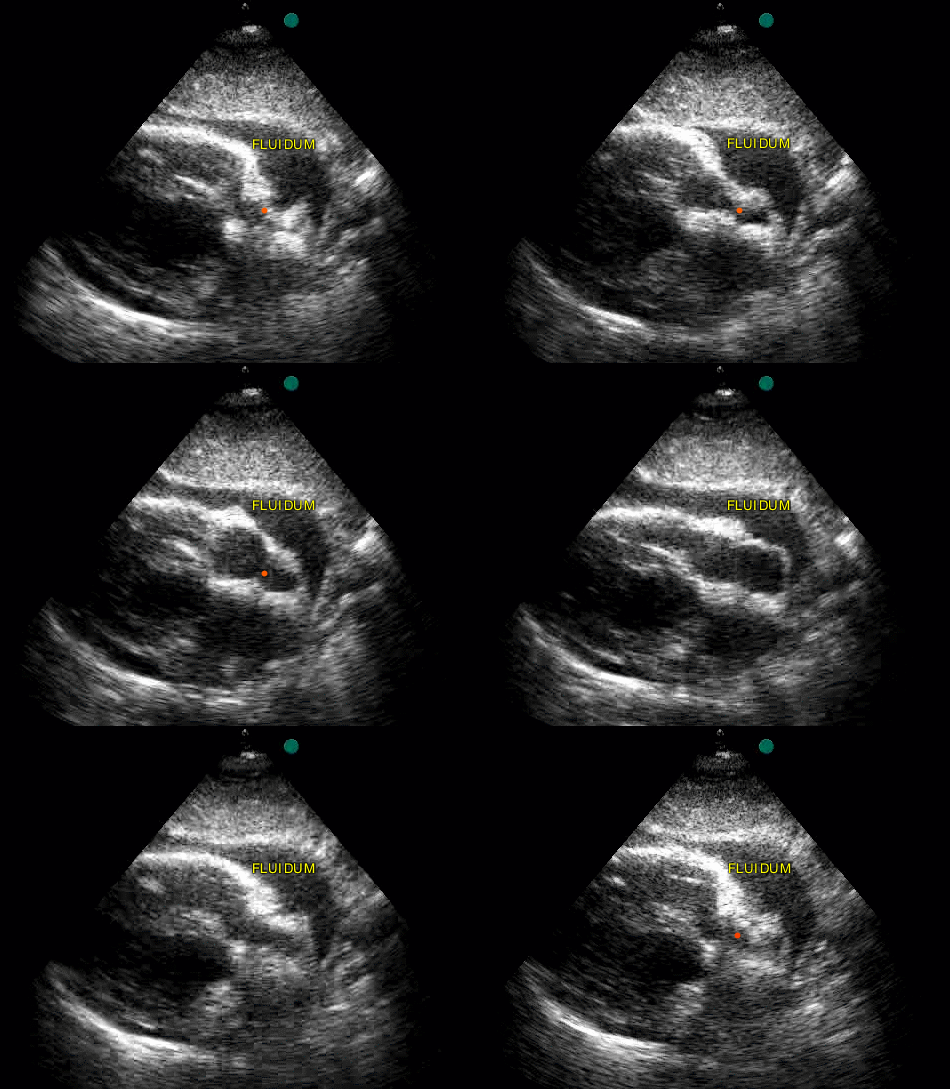
- RUQ (Morison’s pouch): curvilinear probe in the right mid-axillary line around ribs 10–11, marker toward the head. Look between liver and right kidney — and at the inferior tip of the liver, where fluid often appears first.
- LUQ (splenorenal): more posterior and more cranial than the right side. Look above the spleen (subphrenic space) as well as between spleen and kidney.
- Pelvis: just above the pubic symphysis, transverse then longitudinal. Fluid collects behind the bladder (rectovesical pouch or pouch of Douglas).
- Subxiphoid: probe flat under the xiphoid aiming at the left shoulder, looking for a black band of pericardial fluid around the heart.
What a positive FAST looks like
Free fluid is anechoic — pure black — and sits in dependent spaces with sharp, angular edges, unlike the rounded walls of bowel or the gallbladder. A thin black stripe between liver and kidney is a positive RUQ view.
Pitfalls that cause missed fluid
- Too much gain fills fluid with grey and hides small stripes.
- Not sweeping through the whole space — fan the probe from front to back.
- Skipping the liver tip and the subphrenic spaces.
- Treating a negative FAST as “no injury”. It can miss small, early or retroperitoneal bleeds; repeat it and use CT in stable patients.
Real scans
Learn this with your hands. Sonoduck gives you a 3D patient, a probe and a coach that tells you what to do. Free to start, no signup.
Scan your first patientFAQ
How long does a FAST exam take?
About two minutes in experienced hands. It is done at the bedside during resuscitation.
How much fluid can FAST detect?
Commonly quoted thresholds are a few hundred millilitres in Morison’s pouch; sensitivity improves with repeated exams and careful scanning of the liver tip.
Does a negative FAST rule out injury?
No. It does not reliably exclude small or retroperitoneal bleeding or hollow-organ injury.
Related guides
- Morison’s pouch on ultrasoundWhat Morison’s pouch (the hepatorenal space) looks like on ultrasound, how to find it, and how to recognise free fluid — with an interactive simulator.
- Pericardial effusion and tamponade on ultrasoundHow to see a pericardial effusion on bedside echo (subxiphoid and parasternal views) and the ultrasound signs of tamponade. Interactive beating-heart simulator.
- Diagnosing pneumothorax with ultrasoundUltrasound signs of pneumothorax: absent lung sliding, absent B-lines, barcode sign and lung point. Where to scan and the common false positives.
Educational content for learners; follow local protocols and supervision. Simulated images are generated by Sonoduck’s physics model; real images are credited individually.