
Knobology: the four knobs that matter
Machines have dozens of buttons; four matter on almost every scan. Turn them below.
Live simulator — try itFull interactive lesson →
Depth
Start deep to survey, then bring the target to the middle of the screen with a little room behind it.
Gain
Overall brightness. Fluid should be black and solid organs mid-grey. Too much gain fills fluid with fake echoes — the classic reason free fluid gets missed.
Focus
The depth where the beam is narrowest. Place it at or just below what you care about.
Frequency
The highest frequency that still reaches the target: higher is sharper, lower is deeper.
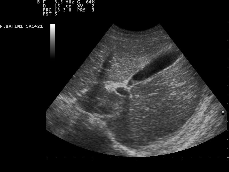
Real scans
Learn this with your hands. Sonoduck gives you a 3D patient, a probe and a coach that tells you what to do. Free to start, no signup.
Scan your first patientFAQ
What is TGC?
Time-gain compensation amplifies later (deeper) echoes so the image is even from top to bottom.
Related guides
- Linear, curvilinear or phased array?The three ultrasound probes explained — frequencies, footprints, image shapes and which to use for vessels, lungs, abdomen and heart — plus the probe marker convention.
- Ultrasound artifacts, explainedAcoustic shadowing, posterior enhancement, reverberation (A-lines), B-lines, mirror image and edge artifacts — why they happen and how they help diagnosis.
- Gallstones and cholecystitis on ultrasoundWhat gallstones look like on ultrasound (bright, shadowing, mobile), the signs of acute cholecystitis, and how to find the gallbladder. Interactive simulator and real scans.
Educational content for learners; follow local protocols and supervision. Simulated images are generated by Sonoduck’s physics model; real images are credited individually.