
Ultrasound artifacts, explained
The machine assumes sound travels straight, at one speed, and bounces once. Artifacts appear when that isn’t true — and many of them are diagnostic.
Live simulator — try itFull interactive lesson →
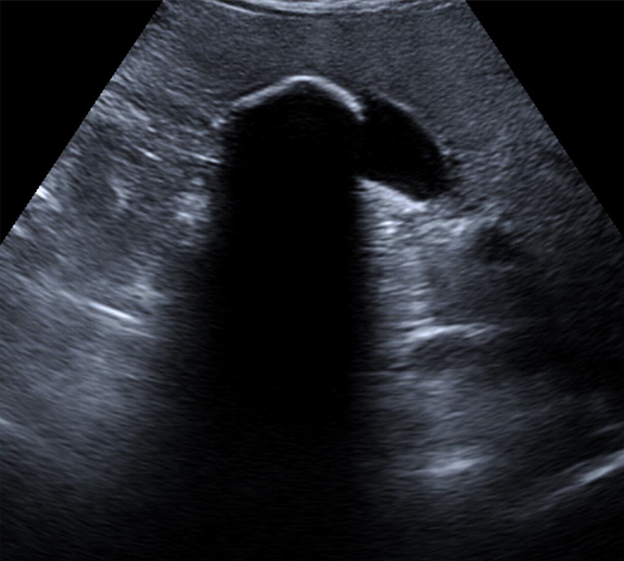
Acoustic shadowing
A dark band behind something that reflects or absorbs almost all sound: stones, bone, calcification. Gas gives a grey, “dirty” shadow.
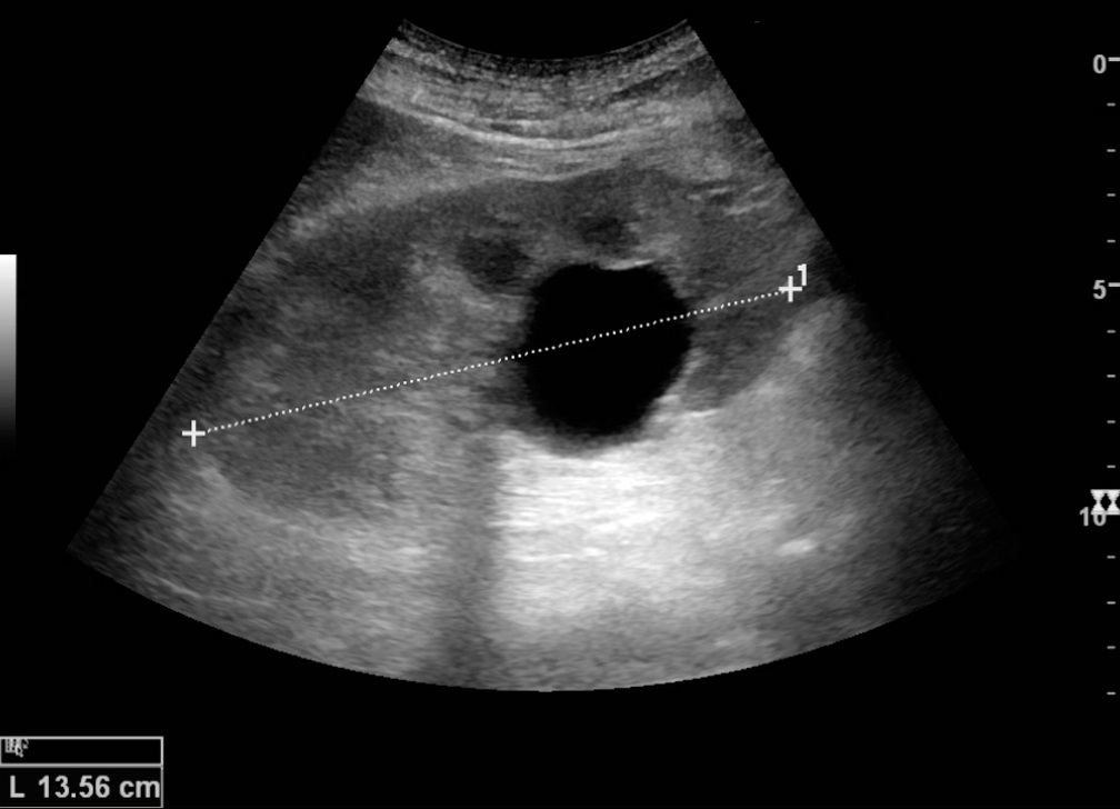
Posterior enhancement
Fluid barely attenuates sound, but the machine still compensates as if it did, so tissue behind fluid looks brighter. It confirms that a black structure is fluid.
Reverberation
Sound bouncing between two strong reflectors appears as evenly spaced copies deeper on the screen — A-lines in the lung, or the ladder behind a needle.
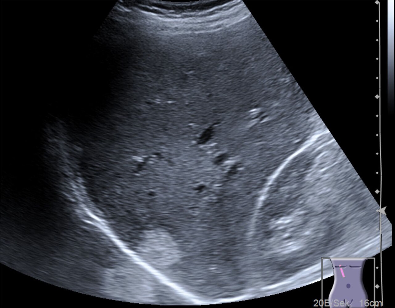
Mirror image
A strong curved reflector such as the diaphragm–lung interface duplicates structures on the far side: liver appearing “above” the diaphragm is normal.
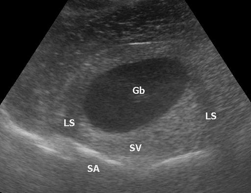
Edge shadows
Thin dark lines from the curved sides of round fluid-filled structures, caused by refraction. Harmless — not stones.
Real scans
Learn this with your hands. Sonoduck gives you a 3D patient, a probe and a coach that tells you what to do. Free to start, no signup.
Scan your first patientFAQ
Are artifacts always bad?
No. Shadowing diagnoses stones, enhancement proves a cyst, and lung ultrasound is built entirely on artifacts.
Related guides
- A-lines vs B-linesThe difference between A-lines and B-lines on lung ultrasound, how many B-lines are abnormal, and what they mean — with real images and a live simulator.
- Gallstones and cholecystitis on ultrasoundWhat gallstones look like on ultrasound (bright, shadowing, mobile), the signs of acute cholecystitis, and how to find the gallbladder. Interactive simulator and real scans.
- Knobology: the four knobs that matterHow to set ultrasound gain, depth, focus and frequency — with a live simulator so you can turn the knobs and see what happens.
Educational content for learners; follow local protocols and supervision. Simulated images are generated by Sonoduck’s physics model; real images are credited individually.