
Gallstones and cholecystitis on ultrasound
The gallbladder sits under the liver just below the right costal margin. Ultrasound is the first-line test for gallstones and acute cholecystitis.
Live simulator — try itFull interactive lesson →
Finding it
Curvilinear probe, subcostal on the right, beam aimed up under the ribs. Ask the patient to breathe in and hold to bring the liver and gallbladder down. Rotate until the black pear-shaped gallbladder is longest, then fan through all of it.
Stone vs polyp
A stone is bright, sits in the dependent part, casts a clean acoustic shadow and moves when the patient rolls. A polyp is fixed to the wall and does not shadow.
Acute cholecystitis
Stones (especially impacted in the neck) plus wall thickening > 3 mm, pericholecystic fluid and a sonographic Murphy’s sign. Wall thickening alone is non-specific — meals, liver disease and heart failure thicken it too.
Real scans
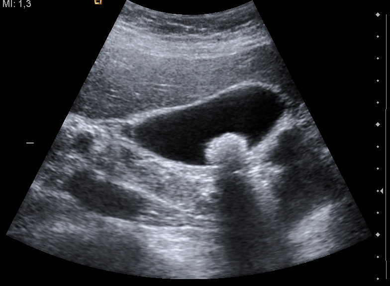
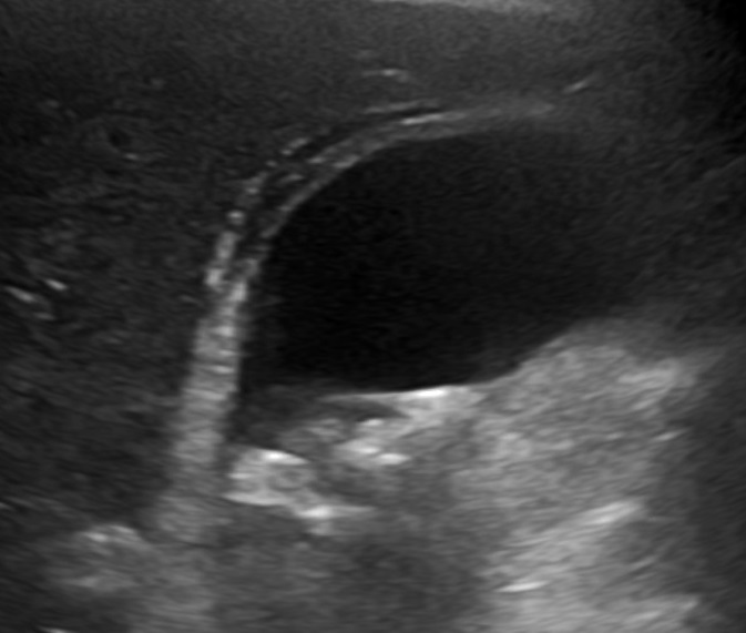
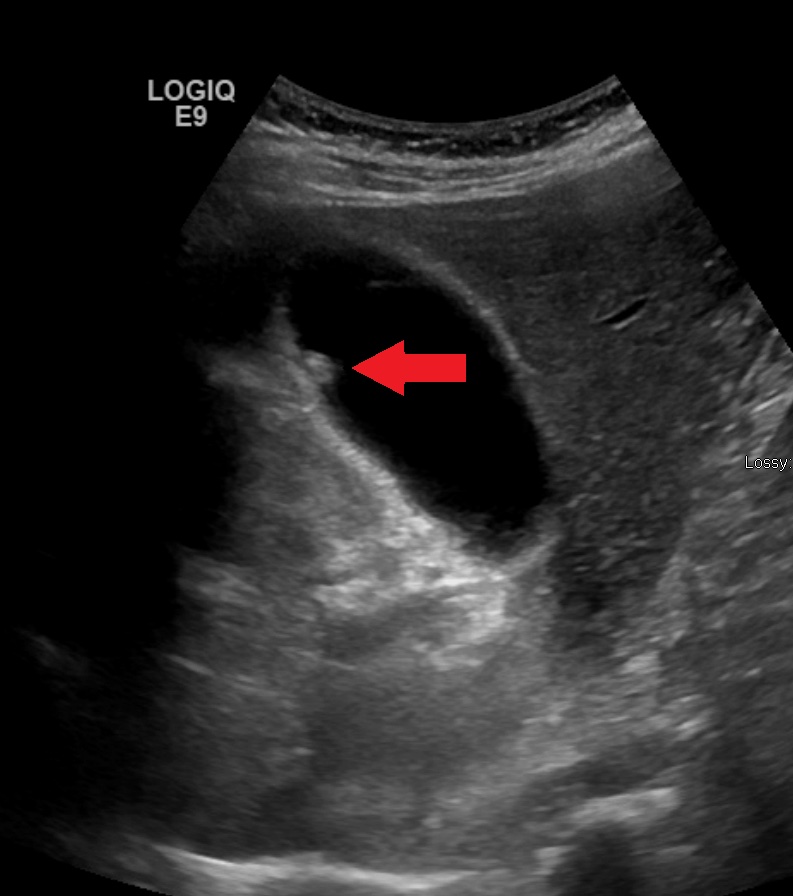
Learn this with your hands. Sonoduck gives you a 3D patient, a probe and a coach that tells you what to do. Free to start, no signup.
Scan your first patientFAQ
Why do gallstones cast a shadow?
They reflect and absorb almost all of the sound, so nothing returns from behind them.
What is the sonographic Murphy’s sign?
Maximal tenderness when pressing the probe directly over the gallbladder.
Related guides
- Ultrasound artifacts, explainedAcoustic shadowing, posterior enhancement, reverberation (A-lines), B-lines, mirror image and edge artifacts — why they happen and how they help diagnosis.
- Knobology: the four knobs that matterHow to set ultrasound gain, depth, focus and frequency — with a live simulator so you can turn the knobs and see what happens.
- Morison’s pouch on ultrasoundWhat Morison’s pouch (the hepatorenal space) looks like on ultrasound, how to find it, and how to recognise free fluid — with an interactive simulator.
Educational content for learners; follow local protocols and supervision. Simulated images are generated by Sonoduck’s physics model; real images are credited individually.