
Morison’s pouch on ultrasound
Morison’s pouch is the potential space between the liver and the right kidney. In a supine patient it is the most dependent part of the upper abdomen, so free fluid collects here early — which makes it the single most important FAST window.
Live simulator — try itFull interactive lesson →
How to get the view
Use a curvilinear probe in the right mid-axillary line around ribs 10–11 with the marker toward the head. Slide down until the liver sits on top and the kidney appears as a bean below it, then fan front-to-back through the whole space. Ask the patient to breathe in if ribs are in the way.
Normal
Liver and kidney capsule touch: you see a thin bright line between them and no black stripe. The diaphragm curves on the head side, with a mirror image of liver texture above it (normal aerated lung).
Positive
An anechoic stripe separating liver from kidney, often extending to the liver tip. In trauma with hypotension, assume it is blood.
Real scans
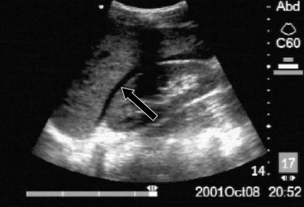
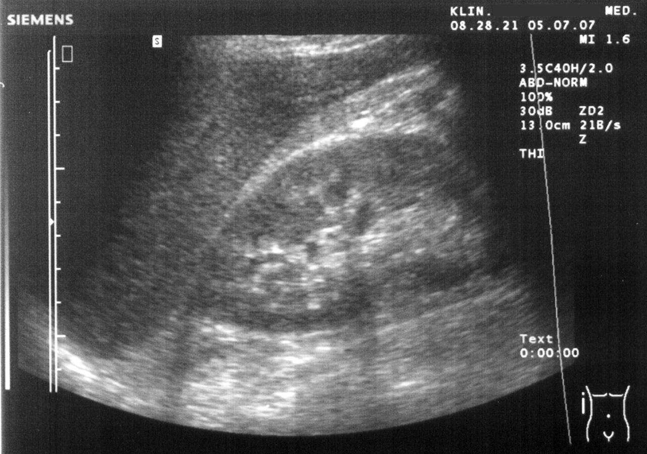
Learn this with your hands. Sonoduck gives you a 3D patient, a probe and a coach that tells you what to do. Free to start, no signup.
Scan your first patientFAQ
Where is Morison’s pouch?
Between the posterior surface of the right lobe of the liver and the right kidney — the hepatorenal recess.
Why does fluid collect there?
It is the most dependent upper-abdominal space when a patient lies flat.
Related guides
- The FAST exam, explained by actually doing itHow to perform a FAST exam: RUQ, LUQ, pelvic and subxiphoid views, what a positive scan looks like, common pitfalls — with a live ultrasound simulator you can try.
- Hydronephrosis on ultrasoundWhat hydronephrosis looks like on ultrasound, how it differs from renal cysts, and how to use the bladder to find the cause. Live simulator and real images.
- Pleural effusion on ultrasoundHow to find a pleural effusion with ultrasound: the flank view, anechoic fluid above the diaphragm, the spine sign and floating lung. Live simulator and real clips.
Educational content for learners; follow local protocols and supervision. Simulated images are generated by Sonoduck’s physics model; real images are credited individually.