
Pericardial effusion and tamponade on ultrasound
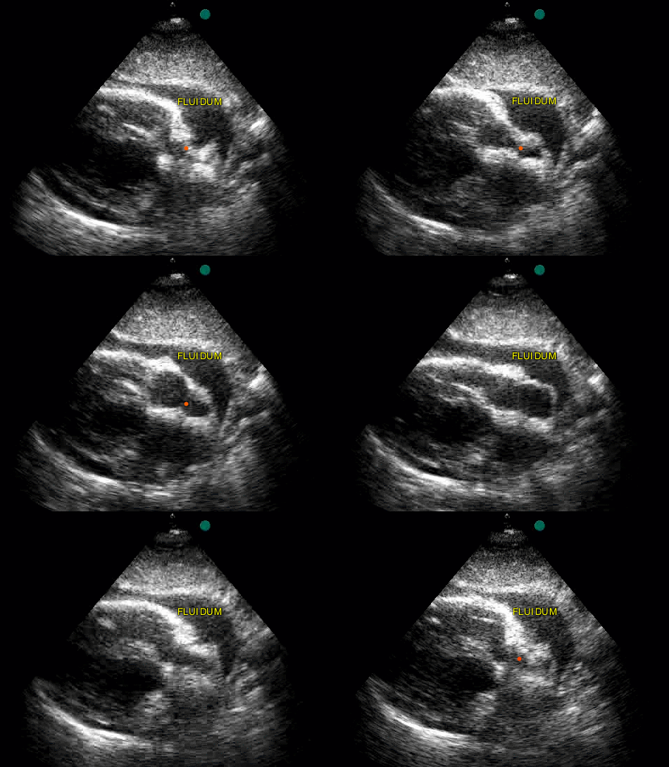
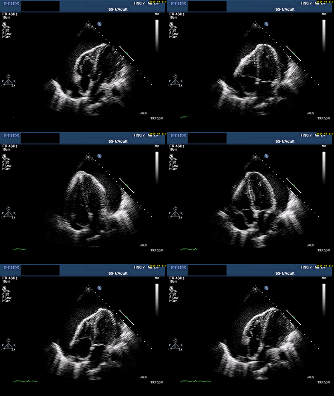
A pericardial effusion is an anechoic band between the bright pericardium and the heart. The question that matters at the bedside is whether it is causing tamponade.
Live simulator — try itFull interactive lesson →
Views
The subxiphoid view looks through the liver at all four chambers — the classic FAST cardiac window. In parasternal long axis, pericardial fluid sits in front of the descending aorta; pleural fluid tracks behind it.
Tamponade signs
- Effusion (size matters less than how fast it accumulated).
- Right atrial collapse in systole and right ventricular collapse in diastole.
- A plethoric IVC that barely changes with breathing.
Real scans
Learn this with your hands. Sonoduck gives you a 3D patient, a probe and a coach that tells you what to do. Free to start, no signup.
Scan your first patientFAQ
How do you tell pericardial from pleural effusion?
In PLAX, pericardial fluid lies anterior to the descending aorta; pleural fluid extends posterior to it.
Can a large effusion be well tolerated?
Yes, if it accumulated slowly. A small, rapid effusion can cause tamponade.
Related guides
- The parasternal long axis viewHow to obtain the parasternal long axis (PLAX) echo view, probe position and orientation, and the structures you should identify — with a live beating-heart simulator.
- The IVC on bedside ultrasoundHow to scan the inferior vena cava, what IVC size and respiratory collapse suggest about right atrial pressure, and the big caveats.
- The FAST exam, explained by actually doing itHow to perform a FAST exam: RUQ, LUQ, pelvic and subxiphoid views, what a positive scan looks like, common pitfalls — with a live ultrasound simulator you can try.
Educational content for learners; follow local protocols and supervision. Simulated images are generated by Sonoduck’s physics model; real images are credited individually.