
Diagnosing pneumothorax with ultrasound
Ultrasound is more sensitive than a supine chest X-ray for pneumothorax. Air rises, so in a patient lying flat you scan the anterior chest first.
Live simulator — try itFull interactive lesson →
The signs, in order
- Absent lung sliding at the pleural line.
- No B-lines (a single B-line rules out pneumothorax at that spot).
- Barcode sign in M-mode instead of the seashore sign.
- Lung point: the edge where sliding lung meets the pneumothorax — highly specific.
False positives
Absent sliding also occurs with apnoea, right mainstem intubation (on the left), pleurodesis or adhesions, and severe ARDS or COPD. Look for a lung point before committing in a stable patient.
Real scans

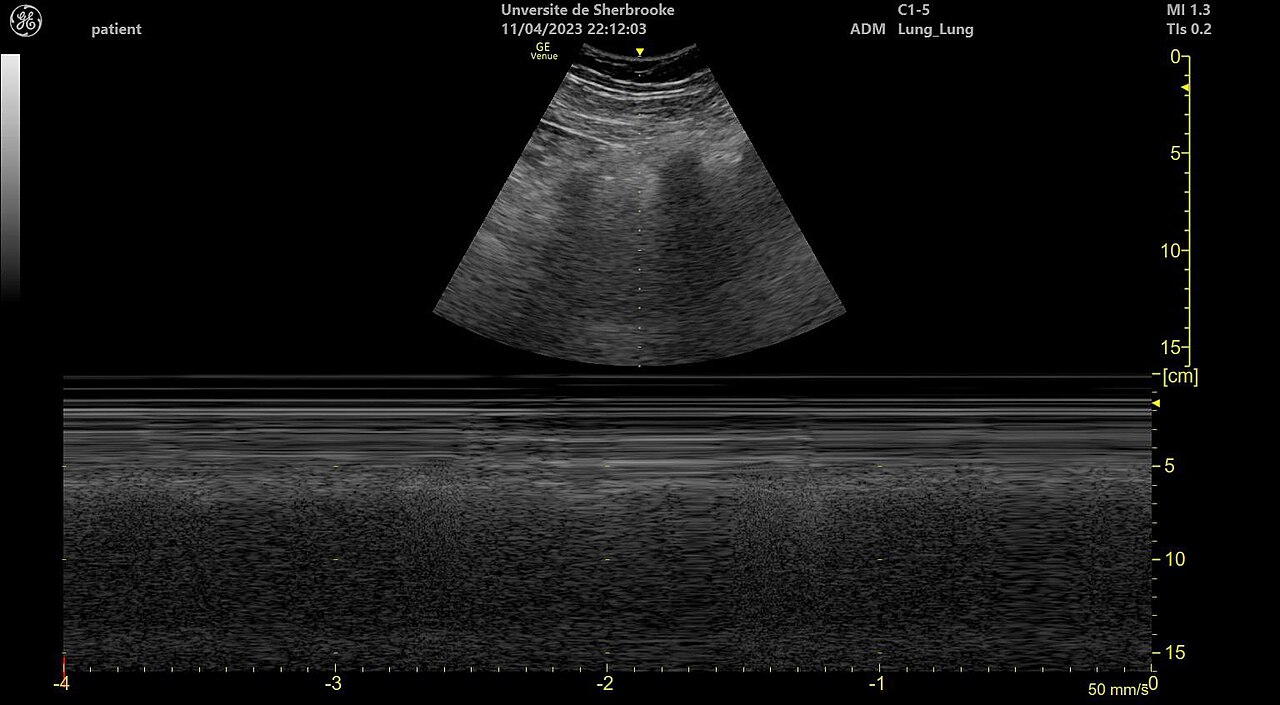
Learn this with your hands. Sonoduck gives you a 3D patient, a probe and a coach that tells you what to do. Free to start, no signup.
Scan your first patientFAQ
Can ultrasound size a pneumothorax?
Roughly — the more lateral the lung point, the larger the pneumothorax — but not precisely.
Which probe should I use?
Linear for the anterior pleural line; curvilinear is fine too.
Related guides
- Lung sliding on ultrasoundWhat lung sliding is, how to see it with a linear probe, the bat sign, seashore sign, and why absent sliding suggests pneumothorax. Try it in a live simulator.
- A-lines vs B-linesThe difference between A-lines and B-lines on lung ultrasound, how many B-lines are abnormal, and what they mean — with real images and a live simulator.
- The FAST exam, explained by actually doing itHow to perform a FAST exam: RUQ, LUQ, pelvic and subxiphoid views, what a positive scan looks like, common pitfalls — with a live ultrasound simulator you can try.
Educational content for learners; follow local protocols and supervision. Simulated images are generated by Sonoduck’s physics model; real images are credited individually.