
Two-point compression ultrasound for DVT
The most learnable test in ultrasound: a normal vein flattens completely when you press. A vein full of clot does not.
Live simulator — try itFull interactive lesson →
Where
1. Groin: the common femoral vein near the saphenofemoral junction. 2. Behind the knee: the popliteal vein. Use a linear probe in transverse.
How
From lateral to medial in the groin: Nerve, Artery, Vein (NAVeL). Press until the artery just starts to deform. If the vein disappears completely, there is no clot at that point. Step along and repeat.
Pitfalls
- Pressing too gently — the commonest cause of a false positive.
- Expecting clot to look grey: fresh clot can be anechoic. Trust compression, not appearance.
Real scans
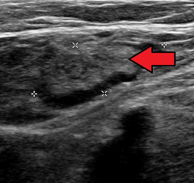
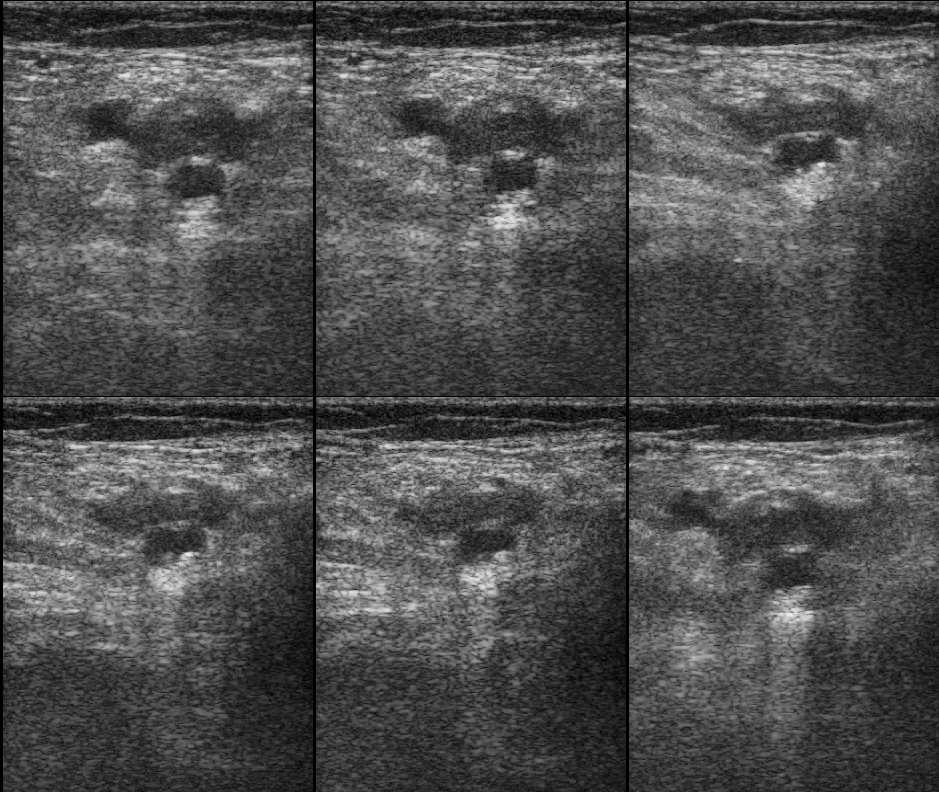
Learn this with your hands. Sonoduck gives you a 3D patient, a probe and a coach that tells you what to do. Free to start, no signup.
Scan your first patientFAQ
Is colour Doppler needed for DVT?
Compression is the primary test; Doppler is an adjunct.
Related guides
- Linear, curvilinear or phased array?The three ultrasound probes explained — frequencies, footprints, image shapes and which to use for vessels, lungs, abdomen and heart — plus the probe marker convention.
- The IVC on bedside ultrasoundHow to scan the inferior vena cava, what IVC size and respiratory collapse suggest about right atrial pressure, and the big caveats.
- Knobology: the four knobs that matterHow to set ultrasound gain, depth, focus and frequency — with a live simulator so you can turn the knobs and see what happens.
Educational content for learners; follow local protocols and supervision. Simulated images are generated by Sonoduck’s physics model; real images are credited individually.