
The IVC on bedside ultrasound
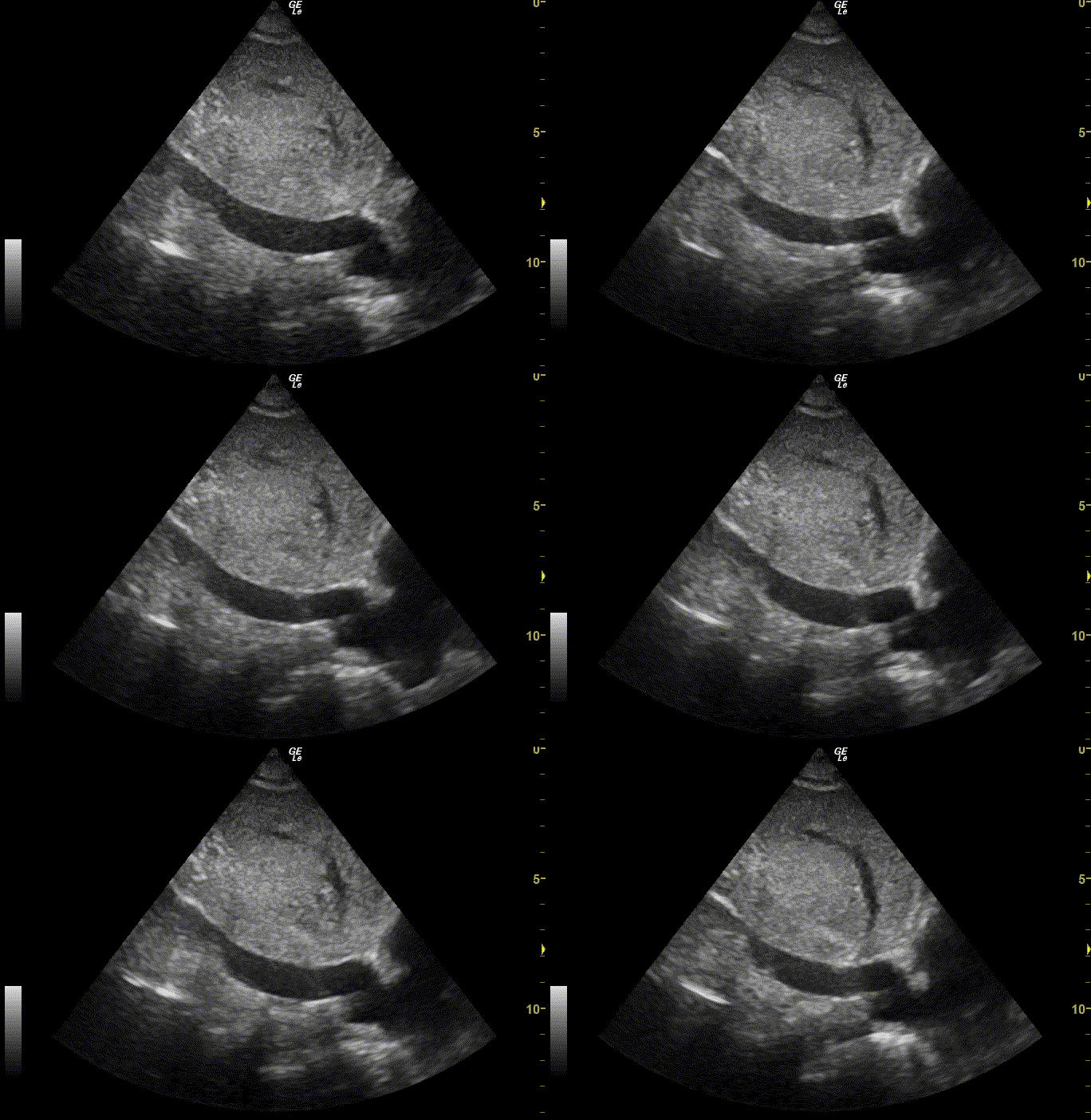
The IVC drains into the right atrium; its size and how much it collapses with a sniff give a rough estimate of right atrial pressure.
Live simulator — try itFull interactive lesson →
How to scan
Curvilinear or phased-array probe, subxiphoid, longitudinal, just right of midline. Follow the IVC into the right atrium; measure about 2 cm from the junction.
Interpretation (ASE)
IVC < 2.1 cm collapsing > 50% with a sniff → low RA pressure (~3 mmHg). IVC > 2.1 cm collapsing < 50% → high RA pressure (~15 mmHg).
Caveats
Mechanical ventilation, raised abdominal pressure, athletes and severe tricuspid regurgitation all confound it. Use it as one clue, not a verdict.
Real scans
Learn this with your hands. Sonoduck gives you a 3D patient, a probe and a coach that tells you what to do. Free to start, no signup.
Scan your first patientFAQ
Does IVC size predict fluid responsiveness?
Only weakly, especially in spontaneously breathing patients.
Related guides
- Pericardial effusion and tamponade on ultrasoundHow to see a pericardial effusion on bedside echo (subxiphoid and parasternal views) and the ultrasound signs of tamponade. Interactive beating-heart simulator.
- Abdominal aortic aneurysm on ultrasoundHow to scan the abdominal aorta at the bedside, how to measure it (outer wall to outer wall), and when it’s an aneurysm. Try the calipers in a live simulator.
- The parasternal long axis viewHow to obtain the parasternal long axis (PLAX) echo view, probe position and orientation, and the structures you should identify — with a live beating-heart simulator.
Educational content for learners; follow local protocols and supervision. Simulated images are generated by Sonoduck’s physics model; real images are credited individually.