
Measuring bladder volume with ultrasound
Urinary retention is a quick bedside diagnosis: measure the bladder in two planes and multiply.
Live simulator — try itFull interactive lesson →
How
Curvilinear probe just above the pubic symphysis. In transverse, measure width and depth (anteroposterior); rotate to longitudinal for length.
Formula
Volume ≈ length × width × height × 0.52 (ellipsoid). Some devices use coefficients up to ~0.7.
Interpretation
A post-void residual above roughly 100–200 mL is abnormal; several hundred millilitres in someone unable to void suggests retention. Check both kidneys for hydronephrosis.
Real scans
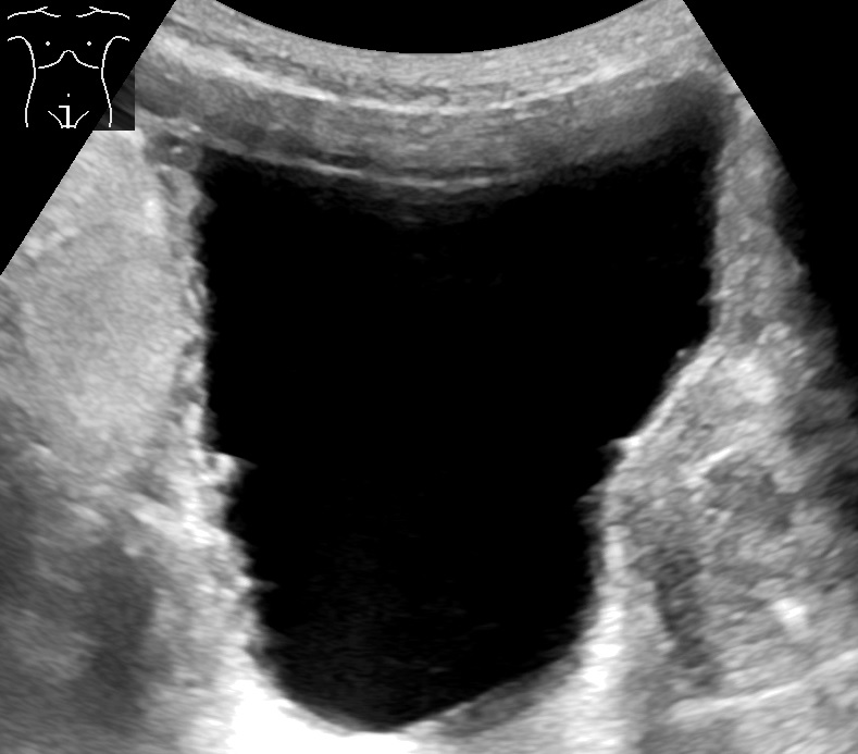
Learn this with your hands. Sonoduck gives you a 3D patient, a probe and a coach that tells you what to do. Free to start, no signup.
Scan your first patientFAQ
Why 0.52?
It is π/6, the volume of an ellipsoid relative to the box enclosing it.
Related guides
- Hydronephrosis on ultrasoundWhat hydronephrosis looks like on ultrasound, how it differs from renal cysts, and how to use the bladder to find the cause. Live simulator and real images.
- Linear, curvilinear or phased array?The three ultrasound probes explained — frequencies, footprints, image shapes and which to use for vessels, lungs, abdomen and heart — plus the probe marker convention.
- The FAST exam, explained by actually doing itHow to perform a FAST exam: RUQ, LUQ, pelvic and subxiphoid views, what a positive scan looks like, common pitfalls — with a live ultrasound simulator you can try.
Educational content for learners; follow local protocols and supervision. Simulated images are generated by Sonoduck’s physics model; real images are credited individually.