
The apical four-chamber view
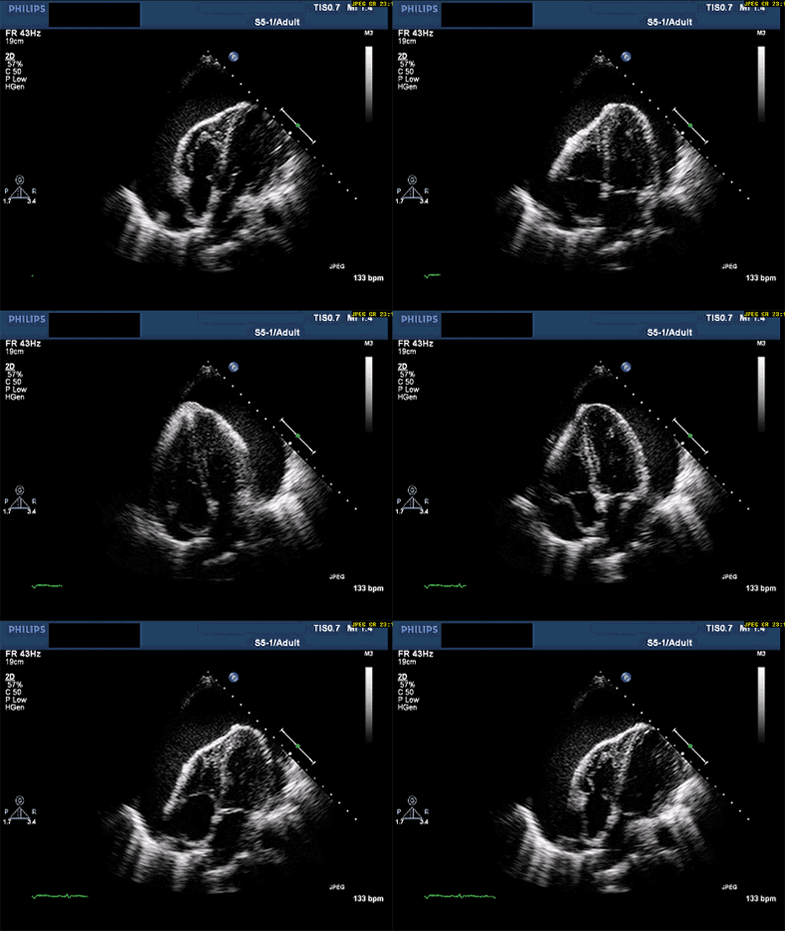
The apical four-chamber view looks up through the apex of the heart and shows all four chambers at once — ideal for comparing the size of the right and left ventricles.
Live simulator — try itFull interactive lesson →
Probe position
Find the apex beat (often below and lateral to the left nipple), phased-array probe with the beam aimed toward the right shoulder. Rolling the patient into the left lateral position brings the heart closer.
What to assess
- RV:LV ratio: the RV is normally under about two-thirds of the LV size; a dilated RV suggests pressure or volume load (e.g., pulmonary embolism).
- Global LV function and regional wall motion.
- Effusion around the free walls.
Real scans
Learn this with your hands. Sonoduck gives you a 3D patient, a probe and a coach that tells you what to do. Free to start, no signup.
Scan your first patientFAQ
Why is the A4C view sometimes foreshortened?
The probe is too high or too medial, cutting through the LV obliquely so the apex looks rounded and the chamber too short.
Related guides
- The parasternal long axis viewHow to obtain the parasternal long axis (PLAX) echo view, probe position and orientation, and the structures you should identify — with a live beating-heart simulator.
- Pericardial effusion and tamponade on ultrasoundHow to see a pericardial effusion on bedside echo (subxiphoid and parasternal views) and the ultrasound signs of tamponade. Interactive beating-heart simulator.
- The IVC on bedside ultrasoundHow to scan the inferior vena cava, what IVC size and respiratory collapse suggest about right atrial pressure, and the big caveats.
Educational content for learners; follow local protocols and supervision. Simulated images are generated by Sonoduck’s physics model; real images are credited individually.